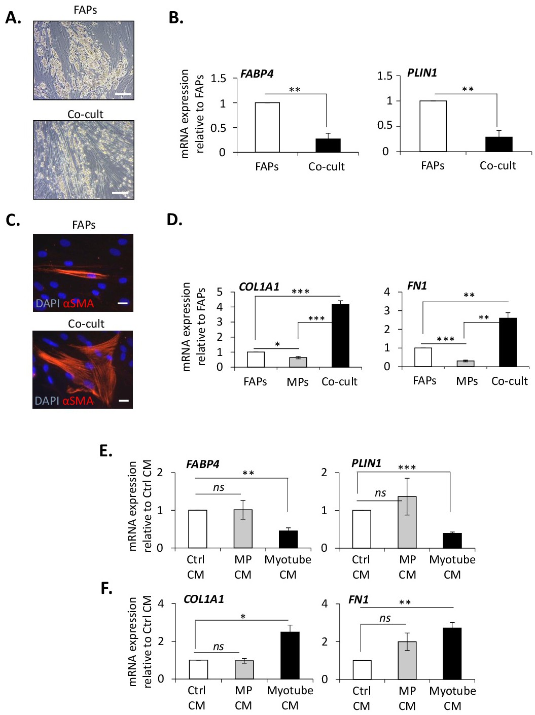
Fig. 3. Myotubes inhibit adipogenesis and stimulate fibrogenesis of FAPs. (A to D) For co-culture assays, FAPs were cultured alone (FAPs) or with MPs (Co-cult) for 10 days in differentiation medium. (A) FAP-derived adipocytes were visualized with phase-contrast microscopy (scale bar: 20 µm). (C) FAP-derived myofibroblasts were visualized by αSMA immuno-labelling. Nuclei were labelled with DAPI (scale bar: 5 µm). Expression of the adipogenic genes FABP4 and PLIN1 (B) and expression of the fibrogenic genes COL1A1 and FN1 (D) were measured by quantitative Q-PCR (n=6 donors). (E and F) For conditioned medium experiments, FAPs were cultured in the presence of conditioned medium from control (Ctrl CM), MPs (MP CM) or differentiated myotubes (Myotube CM) for 10 days. Expression of adipogenic genes (E) and fibrogenic genes (F) was measured by quantitative Q-PCR (n=4 donors). *** P<0.001; ** P<0.01; * P<0.05; ns= non-significant.